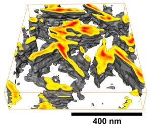
Semi-crystalline elastomeric polypropylene
Our latest result reveals the complex shape and spatial arrangement of
crystalline regions in elastomeric polypropylene. Non-linear image registration
was used to obtain a high image quality over the entire imaging area. This work
was supported by the VolkswagenStiftung. [From: N. Rehse et al., Advanced
Materials; http://dx.doi.org/10.1002/adma.200401473; (c) 2005 by Wiley-VCH.] See also
the related
press release (in German).
Block copolymer microdomain structure
As first example and proof of concept, the microdomain structure of a block copolymer
(polystyrene-block-polybutadiene-block-polystyrene) was
imaged with about 10 nm resolution. The specimen was stepwise eroded by plasma
etching and imaged with tapping mode scanning force microscopy. The left
image is composed from six "slices" and displays the core of a
dislocation within hexagonally ordered cylinders of polystyrene. [From: R. Magerle, Phys.
Rev. Lett. 85, 2749 (2000); (c) 2000 by the American Physical
Society.] Reprint and figures.
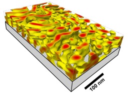
Ultrathin polymer film
Imaging the volume structure of coatings, in particular soft polymeric films on solid substrates, is often a challenge.
In this example, an only about 50 nm thin block copolymer film was imaged from the
original film surface down to the silicon substrate. The layer-by-layer imaging
revealed a novel microdomain structure in the thin film: cylinders with necks. [From: M. Konrad
et al.,
Macromolecules
33, 5518 (2000). (c) 2000 by the American Chemical Society.] Recent
computer simulations showed the underlying physics which causes this pattern
formation in thin films of block copolymers [for details, see: K. S. Lyakhova
et al., J. Chem. Phys. 120,
1127 (2004)].
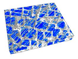
Nickel-based superalloy
Nanotomography of the Nickel-based superalloy CMSX-6. The displayed volume
is about 6.5 x 5.5 x 0.8 µm3 large. Electrochemical etching and
contact mode scanning force microscopy was used to image the material. [From:
M. Göken, R. Magerle, M. Hund, K. Durst, in: Prakt. Metallogr. Sonderbd.
35, Metallographietagung Berlin 2003, (Ed.) P.D. Portella,
Werkstoff-Informationgesellschaft mbH, Frankfurt (2004), S. 257].