Nanotomography R. Magerle, Phys. Rev. Lett. 85, 2749 (2000); 
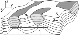
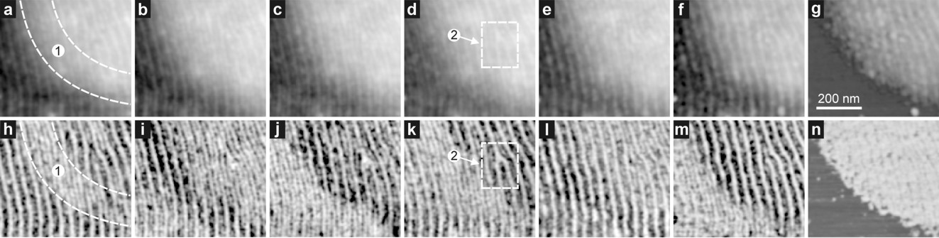
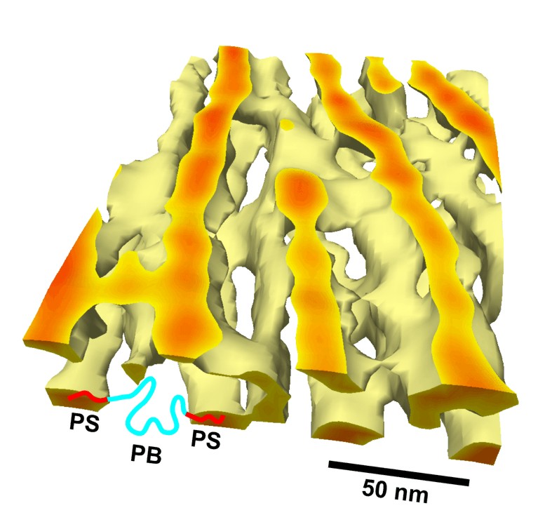
Abstract: Scanning probe microscopy (SPM) can be expanded to volume imaging. As an example, the core of a dislocation within the three-dimensional (3D) spatial microdomain structure of poly(styrene-block-butadiene-block-styrene) was imaged with ~ 10 Reprint: pdf (766 kB) (c) 2000 by the American Physical Society. Permissions to reproduce the figures can be obtained from the author. |
|
|
Fig. 1 Schematic illustration of the principle of volume reconstruction from a series of scanning probe microscopy images. In general the surfaces Sn on which the property P(Sn) is measured are curved. |
|
Fig. 2 Series of TappingModeTM SFM images from a thin SBS film. Topography [(a)-(g)] and the corresponding phase images [(h)-(n)] are shown. Between each pair of images a 7.5±0.2 nm (on average) thick layer has been removed from the sample by plasma etching. For (1), see text. Region (2) is displayed as 3D image in Fig. 3. |
|
Fig. 3 3D image of the isosurface enclosing the volume with normalized phase > 0.11 reconstructed from the series of phase and topography images shown in Fig. 2. This can be interpreted as PS cylinders within a 200 x 160 x 45 nm3 large portion of the SBS film. In the center a branching of a PS cylinder (1) into four other PS cylinders (2)-(5) is displayed which is the core of a dislocation line in the SBS microdomain structure of hexagonally ordered PS cylinders. For size comparison and illustration of the molecular structure of the material a SBS molecule bridging two PS domains is sketched. |
|
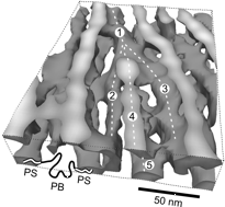
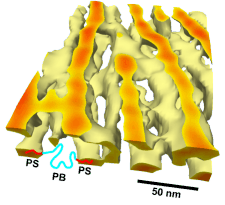
Color version of Fig. 3
|
webmaster@nanotomography.de (08/2005)