|
Most modern materials have complex structures on the micron and nanometer scale. A detailed study of the structure-property relationship of many materials is often hindered, as most microscopy techniques available today are limited to two-dimensional imaging. Nanotomography, a new high-resolution imaging technique based on scanning probe microscopy, might help to solve this problem.
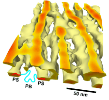
Nanotomography is similar to an excavation on the nanometer scale: With suitable etching or polishing techniques (for instance wet chemical etching, plasma etching or chemo-mechanical polishing) the specimen is eroded step by step and the chemical composition of each freshly exposed surface is imaged with scanning probe microscopy. From the resulting series of images, separated in depth by only a few nanometers, the specimen’s three-dimensional microstructure can be reconstructed. With Nanotomography we have imaged with 10 nm resolution various materials:
-
individual defects in blockcopolymers (see figure),
-
ultrathin films of blockcopolymers,
-
semi-crystalline elastomeric polypropylen,
-
metallic Nickel-based superalloys.
For more details see the image gallery, our publications, or contact me directly. I would be happy to send you reprints and to discuss with you, how your material could be imaged with Nanotomography. Our aim is to explore the new possibilites of high-resolution volume imaging that Nanotomogray offers. With the success of scanning probe microscopy in mind, volume imaging with scanning probe microscopy promises new insights into the physics of condensed matter on a nanometer scale.
Robert Magerle